Welcome to Metamora Equine!
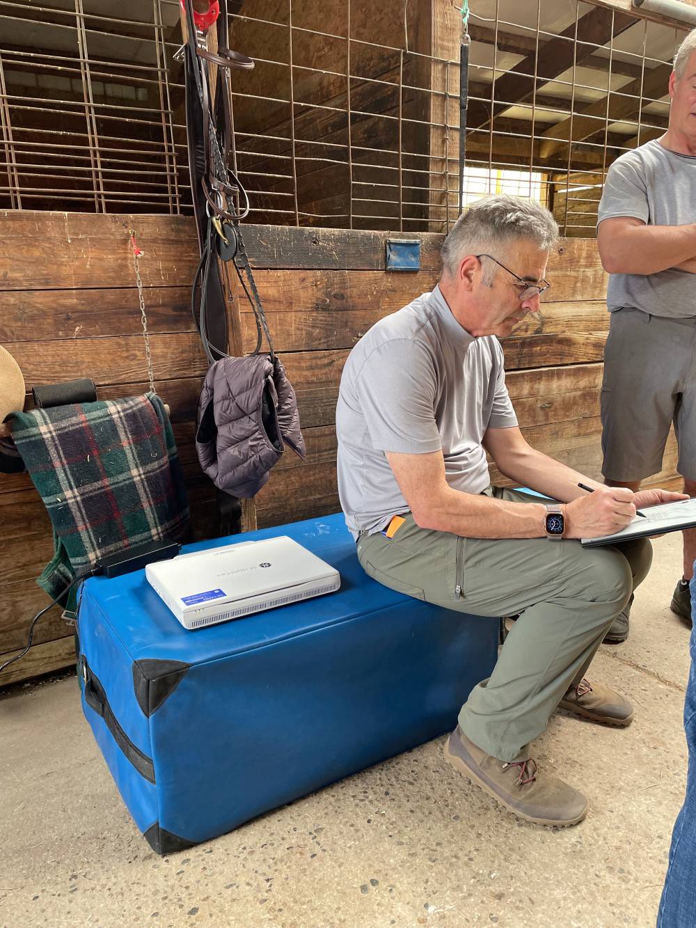
Metamora Equine is a 100% ambulatory practice specializing in advanced sports medicine for competition horses, led by board-certified Dr. Roland Thaler. Based in Davisburg, Michigan, we serve:
- Detroit suburbs
- Columbus and Wilmington, Ohio
- Monroe, North Carolina
- Aiken South Carolina
- Major showgrounds in the Midwest and Southern US.
Specializing in advanced diagnostics, complex lameness cases, pre-purchase consultations, and performance-focused care for elite sport horses, we offer academic-level sports medicine directly at your barn or competition venue. Our thoughtful, thorough approach is rooted in 40 years of experience, to understand the source of discomfort and build a clear, effective care plan. For veterinarians and investors, Metamora Equine (MEPC) is a rare opportunity to acquire a highly profitable, respected mobile practice built on clinical excellence, advanced technology, and client trust.